Physical Characteristics
The horseshoe crab has been described as an armored box that moves. Their appearance is similar to the prehistoric and extinct trilobite. Looking at the exterior of the crab, the body is divided into three sections. These three sections comprise the horseshoe crab's hardened exoskeleton. The exoskeleton is shed periodically as the crab grows.
Prosoma
(cephalothorax) - The largest section of the horseshoe crab. From a top view, it is shaped like a horse's shoe. Several eyes are found on the exterior of the prosoma.
Opisthosoma
(abdomen) - The abdomen is the center section of the shell and attaches to the cephalothorax using a hinge. From a top view, moveable spines are visible along the edge of the abdomen.
Telson
(tail) - The tail is attached to the abdomen at the terminal base. The horseshoe crab uses its telson to steer and right itself if it becomes inverted in the tidal zone. Contrary to popular belief, the tail is not a poisonous stinger. Occasionally, horseshoe crabs are found with a misshapened telson. This is usually due to a physical injury of the telson.
Eyes
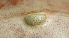
 Horseshoe crabs have a total of 10 eyes used for finding mates and sensing light. The most obvious eyes are the 2 lateral compound eyes. These are used for finding mates during the spawning season. Each compound eye has about 1,000 receptors or ommatidia. The cones and rods of the lateral eyes have a similar structure to those found in human eyes, but are around 100 times larger in size. The ommatidia are adapted to change the way they function by day or night. At night, the lateral eyes are chemically stimulated to greatly increase the sensitivity of each receptor to light. This allows the horseshoe crab to identify other horseshoe crabs in the darkness.
The horseshoe crab has an additional five eyes on the top side of its prosoma. Directly behind each lateral eye is a rudimentary lateral eye. Towards the front of the prosoma is a small ridge with three dark spots. Two are the median eyes and there is one endoparietal eye. Each of these eyes detects ultraviolet (UV) light from the sun and reflected light from the moon. They help the crab follow the lunar cycle. This is important to their spawning period that peaks on the new and full moon. Two ventral eyes are located near the mouth but their function is unknown.
Multiple photoreceptors located on the telson constitute the last eye. These are believed to help the brain synchronize to the cycle of light and darkness.
(Check out a diagram of the horseshoe crab's 10 eyes).
Gills
A horseshoe crab absorbs oxygen from the water using gills that are divided into 5 distinct pairs located under the abdomen. Each pair of gills has a large flap-like structure covering leaf-like membranes called lamellae. Gaseous exchange occurs on the surface of the lamellae as the gills are in motion. Each gill contains approximately 150 lamellae that appear as pages in a book. They are commonly called book gills. The gills also function as paddles to propel juvenile horseshoe crabs through the water.
Mouth & Legs
The horseshoe crab has 6 pairs of appendages on the underside. The first pair are called Chelicerae which are used to place food in the mouth. The next pair are called pedipalps. These are the first of the 3 sets of ambulatory legs and in males are used for grasping the female. The "last" pair of legs are called the "pusher legs" they have a leaflike structure at the end that is used for locomotion. The pusher leg also has a spatulate organ called the flabellum that is used to test the composition of the water passing to the gills.
The base of each leg is covered with inward pointing spines called gnathobases that move food towards the mouth located between the legs. As the legs are moving, food is crushed and macerated. There are also 2 small chelicera appendages that help guide food into the mouth.
Circulatory System
The horseshoe crab has a developed circulatory system. A long tubular heart runs down the middle of the prosoma and abdomen. The rough outline of the heart is visible on the exoskeleton and at the hinge. Blood flows into the book gills where it is oxygenated in the lamellae of each gill. The flapping movement of the gills circulates blood in and out of the lamellae. Oxygenated blood is returned to the heart for distribution throughout the horseshoe crab.
Male/Female Variations
Several distinct variations between males and females occur in horseshoe crabs. Upon reaching maturity at 9-10 years old, the female horseshoe crab will molt an additional one or two more times. As a result, the female crab is considerably larger than the male. Also, the mature male horseshoe crab will develop a modified first pair of walking legs. The new legs (adapted pedipalps) have a hooklike structure that resembles a boxing glove. The male horseshoe crab uses the modified legs to clasp onto the shell of the female during spawning.
Prior to reaching maturity males and females are identified by the shape of their genital pores. The pores can be found behind the first gill cover at the base of the first pair of book gills. On a male, the genital pores are firm pointed structures and white in color. The female genital pores are broad convex structures similar in appearance to small bumps.
Take a look at some photos to see the differences.
To learn more, visit the links on the left.